

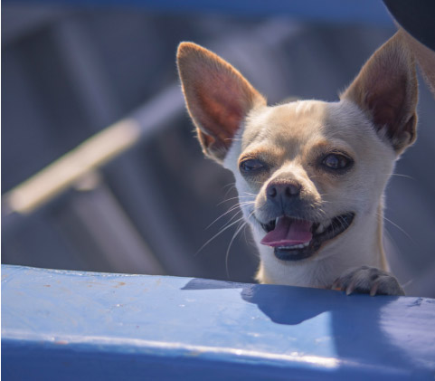
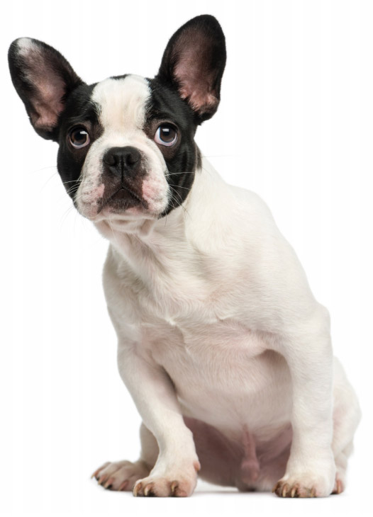
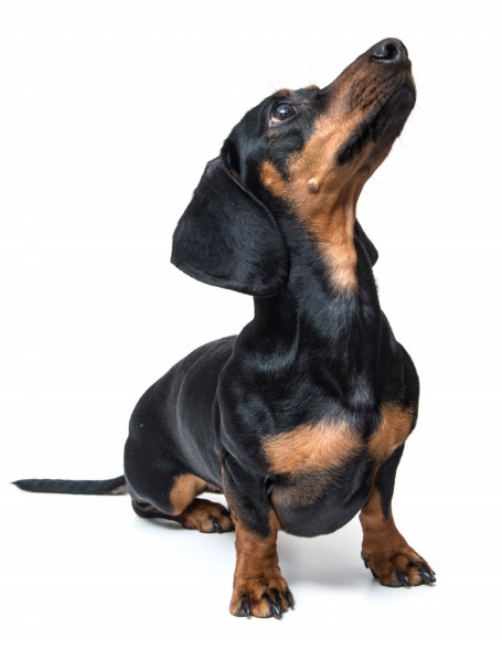
Canine intervertebral disc disease (IVDD) involves degeneration of the intervertebral discs that often leads to disc herniation resulting in spinal cord or spinal nerve compression. The syndrome can affect all breeds but toy breed dogs such as dachshunds, poodles, Pekingese, etc. are more commonly affected. Clinical signs range from intermittent pain to complete paralysis.
The spine is made up of block bones called vertebrae. Dogs have seven cervical (neck), thirteen thoracic (chest), seven lumbar (back), three fused sacral (tail bone) and a variable number of tail vertebrae. Each vertebra is made up of a body, the lamina, which houses the spinal cord, and the facets that are partially responsible for the connection from one vertebra to the next.
Intervertebral discs are situated between each vertebra and in dogs and cats the disc sits just beneath, or ventral to, the spinal canal (figure 1). The discs are comprised of a fibrous outer ring called the annulus fibrosis and an inner gel called the nucleus pulposus and act as both connectors and shock absorbers between each vertebra.
Canine intervertebral disc disease manifests by two different pathologic processes classified as Hansen type one, Hansen type two and less commonly, Hansen type three. In toy breed dogs the discs degenerate and dehydrate (Hansen type one). This process can begin as early as one year of age. As discs lose resiliency and the outer layers weakens, the inner nucleus pulposus is at risk to displace upward (herniate) resulting in spinal cord trauma and compression (figure 2). The herniation often involves a rapid burst of disc material causing acute pain or paralysis. In large breed dogs the pathologic process involves cycles of gradual break down, followed by fibrous repair resulting in disc bulging and insidious or slow onset of clinical signs (Hansen type two). Colorado Canine Orthopedics’ surgeons focus on toy breed dogs and the following discussion refer specifically to Hansen type one disc disease. Disc herniations can also be classified according to location: cervical (neck), thoracolumbar (mid-back) or lumbo-sacral (lower back).
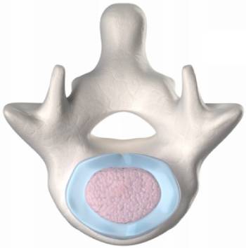
Figure 1: Normal Intervertebral Disc
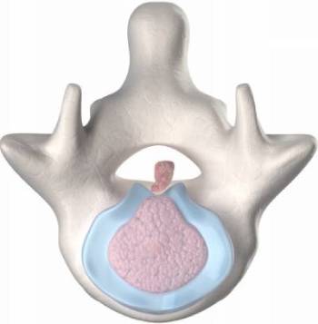
Figure 2: Disc Herniation
Clinical signs associated with Hansen type one (small breed) cervical IVDD vary depending the exact location and force of the disc herniation and the amount of material herniated. Often the clinical signs appear suddenly and include neck pain, crying out when moving or being picked up, and an extremely stiff neck. The head is often held in a downward position and a front limb lameness may be present. Tetraparesis (stumbling on all four limbs) is possible but uncommon. Dogs with cervical IVDD are almost always ambulatory (walking).
Numerous imaging modalities can be used to diagnose cervical IVDD. Plain radiographs (X-rays) are a useful screening tool but usually are not accurate in obtaining a conclusive diagnosis. Computerized tomography (CT) is quite accurate in diagnosing cervical IVDD in small breed dogs (figure 3). Contrast media is often used in conjunction with CT imaging. Magnetic resonance imaging (MRI) is also used to diagnose canine spinal disorders but is most applicable in large breed dogs, lumbosacral IVDD and neoplasia.
The treatment options for cervical IVDD are variable depending on the severity of the clinical signs, duration of clinical signs and number of recurrences. Dogs with mild neck pain should be treated conservatively with cage confinement, acupuncture, possibly corticosteroids and pain medications as needed. In cases where the pain is severe and/or neurologic deficits, are present such as stumbling while walking, surgical decompression is recommended. In cases of multiple recurrences, surgery is also recommended.
Surgery involves making a small opening through the ventral (underside) aspect of the vertebra allowing access to the spinal canal and herniated disc. The actual name of the procedure is ventral slot. Most animals spend one night in ICU and are able go home the following day.
The day following surgery most patients are comfortable and able to return home. Crate confinement with occasional short leash walks is recommended for the first several weeks following surgery (lap sitting is encouraged). Most dogs can return to full function after 4 weeks.
Overall the prognosis is excellent following surgery. Additional subsequent disc herniations are possible and studies suggest the probability is about 15%.
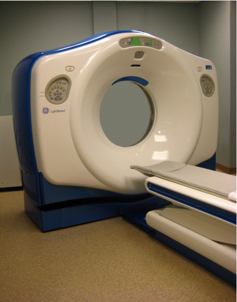
Figure 3: CT Scanner
Clinical signs of thoracolumbar (mid-back) intervertebral IVDD range from pain to complete paralysis and often develop rapidly. In mild cases, dogs have back pain only. In more advanced cases, the patient is ambulatory but staggers on the rear limbs. In more severe cases, paraplegia (inability to use the rear limbs) is present. In the most severe cases paraplegia is present and deep pain sensation is lost. It is not uncommon for the clinical signs to progress from pain only, to paraplegia over 12-24 hours.
Numerous imaging modalities can be used to diagnose thoracolumbar IVDD. Plain radiographs (X-rays) are a useful screening tool but usually are not accurate in obtaining a conclusive diagnosis. Computerized tomography (CT) is quite accurate in diagnosing thoracolumbar IVDD in small breed dogs (figure 4). Contrast media is often used in conjunction with CT imaging. Magnetic resonance imaging (MRI) is also used to diagnose canine spinal disorders but is most applicable in large breed dogs, lumbosacral IVDD and neoplasia.
The treatment options for thoracolumbar IVDD are variable depending on the severity and duration of the clinical signs. Dogs with back pain only, should be treated conservatively with cage confinement, acupuncture, possibly corticosteroids and pain medications as needed. These patients should be closely monitored for progression of clinical signs. In cases where neurologic deficits are present, such as stumbling while walking the decision between conservative management and surgical intervention become somewhat opiniated and vague. If clinical signs are rapidly deteriorating, for example going from pain only to stumbling (paresis) over several hours, surgical decompression should be considered. In animals unable to walk, surgery is recommended within 12-24 hours.
Surgery involves a small surgical approach to spine followed by burring away the lamina (side portion of the vertebra), thereby exposing the spinal cord and herniated disc material. The material is gently removed from the spinal canal. The procedure is called a hemilaminectomy. Most animals spend one night in ICU and are able go home the following day.
The day following surgery most patients are comfortable and able to return home. A urinary catheter is typically left in place for 5-7 days. Crate confinement is recommended for the first several weeks following surgery (lap sitting is encouraged). Use of the hind limbs is often present after two weeks although the return to functional walking may take up to 4 weeks. Most dogs can return to full function after about 6-8 weeks.
The prognosis for non-ambulatory, deep pain present dogs with thoracolumbar IVDD is excellent. In a series of 50 surgical cases followed at Colorado Canine Orthopedics, 98% of dogs that had deep pain sensation at the time of surgery returned to walking. Dogs that have lost deep pain sensation have a less favorable prognosis, and although the exact percentage if not well established, approximately 50 percent of return to some level of functional walking. Additional subsequent disc herniations are possible and studies suggest the probability is about 15%.
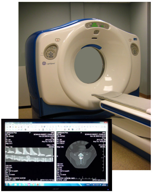
Figure 4: CT scan showing Disc Herniation
Surgical costs for small breed IVDD surgery vary depending on the extent of the injury. We will provide information about costs for your dog’s case at your consultation. Please feel free to contact us with questions.
At Colorado Canine Orthopedics we are committed to providing only state of the art, non-compromised pet healthcare. We realize some pet owners may find this level of care relatively costly. However, despite the inherently expensive nature of our work, we are dedicated to providing the highest level of care at the most affordable price possible. We believe if you compare our fees to other specialty practices you will find this true.
Colorado Canine Orthopedics
5528 North Nevada
Colorado Springs, CO 80918
Tel: 719-264-6666


Call our office today and speak with one of our Pet Care Coordinators to schedule a consultation.
Call our office today and speak with one of our Pet Care Coordinators to schedule a consultation.

Colorado Canine Orthopedics
5528 North Nevada
Colorado Springs, CO 80918
Tel: 719-264-6666